|
| |
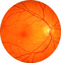
Retinal Vein Occlusion
Retinal vein occlusion
occurs when the circulation of a retinal vein becomes obstructed by an
adjacent blood vessel, causing hemorrhages in the retina. Swelling and
ischemia (lack of oxygen) of the retina as well as glaucoma are fairly
common complications.
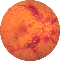
The visual symptoms can vary in severity from one person to the next,
and are dependent on whether the central retinal vein or a branch
retinal vein is involved. Patients who experience a branch vein
occlusion often notice a gradual improvement in their vision as the
hemorrhage resolves. Recovery from a central vein occlusion is much less
likely.
SIGNS AND SYMPTOMS
•Sudden onset
•Blurred or missing area of vision (if a branch vein is involved)
•Severe loss of central vision (if a central vein is involved)
•More
common after age 60 (males and females)
DETECTION AND DIAGNOSIS
Vein occlusion is diagnosed by examining the retina with an
ophthalmoscope. Fluorescein angiography may be performed in some cases
to study the circulation of the retina and to determine the extent of
macular edema or swelling.
TREATMENT
Following a vein
occlusion, the primary concern is to treat the secondary complications.
If areas of the retina are oxygen-deprived, LASER may be used to prevent
growth of delicate vessels that could break, bleed or cause glaucoma.
The following are common risk factors for vein occlusion:
•Diabetes
•Hypertension
•Cardiovascular disease
|
|